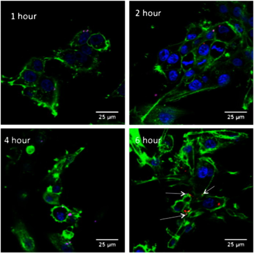
Confocal microscopy provides only a marginal improvement in both axial ( z along the optical axis) and lateral ( x and y in the specimen plane) optical resolution, but is able to exclude secondary fluorescence in areas removed from the focal plane from resulting images. The problem is compounded by thicker specimens (greater than 2 micrometers), which usually exhibit such a high degree of fluorescence emission that most of the fine detail is lost. In a conventional widefield optical epi-fluorescence microscope, secondary fluorescence emitted by the specimen often occurs through the excited volume and obscures resolution of features that lie in the objective focal plane. In fact, confocal technology is proving to be one of the most important advances ever achieved in optical microscopy. There has been a tremendous explosion in the popularity of confocal microscopy in recent years, due in part to the relative ease with which extremely high-quality images can be obtained from specimens prepared for conventional fluorescence microscopy, and the growing number of applications in cell biology that rely on imaging both fixed and living cells and tissues.

The basic key to the confocal approach is the use of spatial filtering techniques to eliminate out-of-focus light or glare in specimens whose thickness exceeds the immediate plane of focus.
#Snapgene confocal microscopy serial
Confocal microscopy offers several advantages over conventional widefield optical microscopy, including the ability to control depth of field, elimination or reduction of background information away from the focal plane (that leads to image degradation), and the capability to collect serial optical sections from thick specimens.


 0 kommentar(er)
